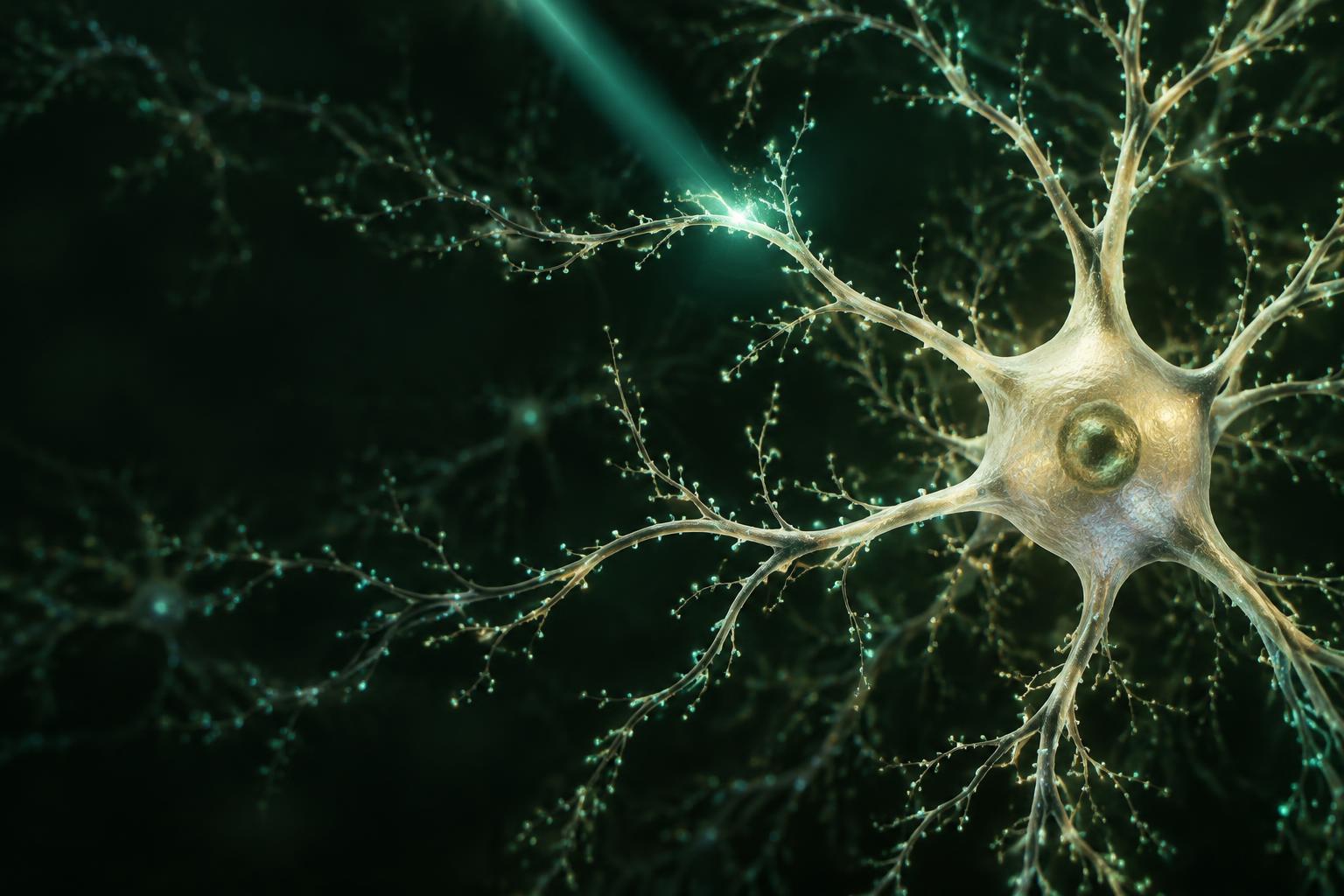
The brain doesn’t reveal its secrets easily. Watching neurons fire can tell us what happens during a thought or a movement. Finding out why it happens is a much harder question.
That is where optogenetics changed the conversation. By making selected cells responsive to light, researchers can switch specific parts of a neural circuit on or off and observe what follows. Karl Deisseroth helped turn that idea into a practical tool for neuroscience.
If you are searching for Karl Deisseroth optogenetics brain disorders, the essential distinction is this: the research helps explain how brain circuits contribute to symptoms. It does not mean that shining light on the head—or taking a supplement—provides an optogenetic treatment.
Optogenetics combines light and genetic tools to study precisely targeted cells. Its findings are important for brain-disorder research, but an experimental result in an animal is not the same as an established treatment for a person.
Who is Karl Deisseroth?
Karl Deisseroth is a Stanford psychiatrist and neuroscientist whose work connects questions about mental health with the biology of neural circuits. He is a pioneering contributor to optogenetics, a field built through the efforts of many researchers—not a single discovery made in isolation.
A landmark 2005 paper by Edward Boyden, Feng Zhang, Ernst Bamberg, Georg Nagel and Deisseroth demonstrated rapid optical control of neuronal activity using a light-sensitive microbial protein. The study helped establish a way to manipulate neurons with millisecond-scale timing. [1]
The importance of that precision is easy to miss. A brain circuit is not simply a region that is “active” or “inactive.” It contains different cells, connections and timing patterns. Optogenetics gives researchers a way to test the contribution of selected elements rather than treating the entire circuit as one undifferentiated block.
How does optogenetics work?
The name joins two ingredients: optics, or light, and genetics, the means of making particular cells light-sensitive. Many optogenetic tools use proteins called opsins. Depending on the protein and experimental conditions, illumination can encourage a neuron to fire or suppress its activity. [2]
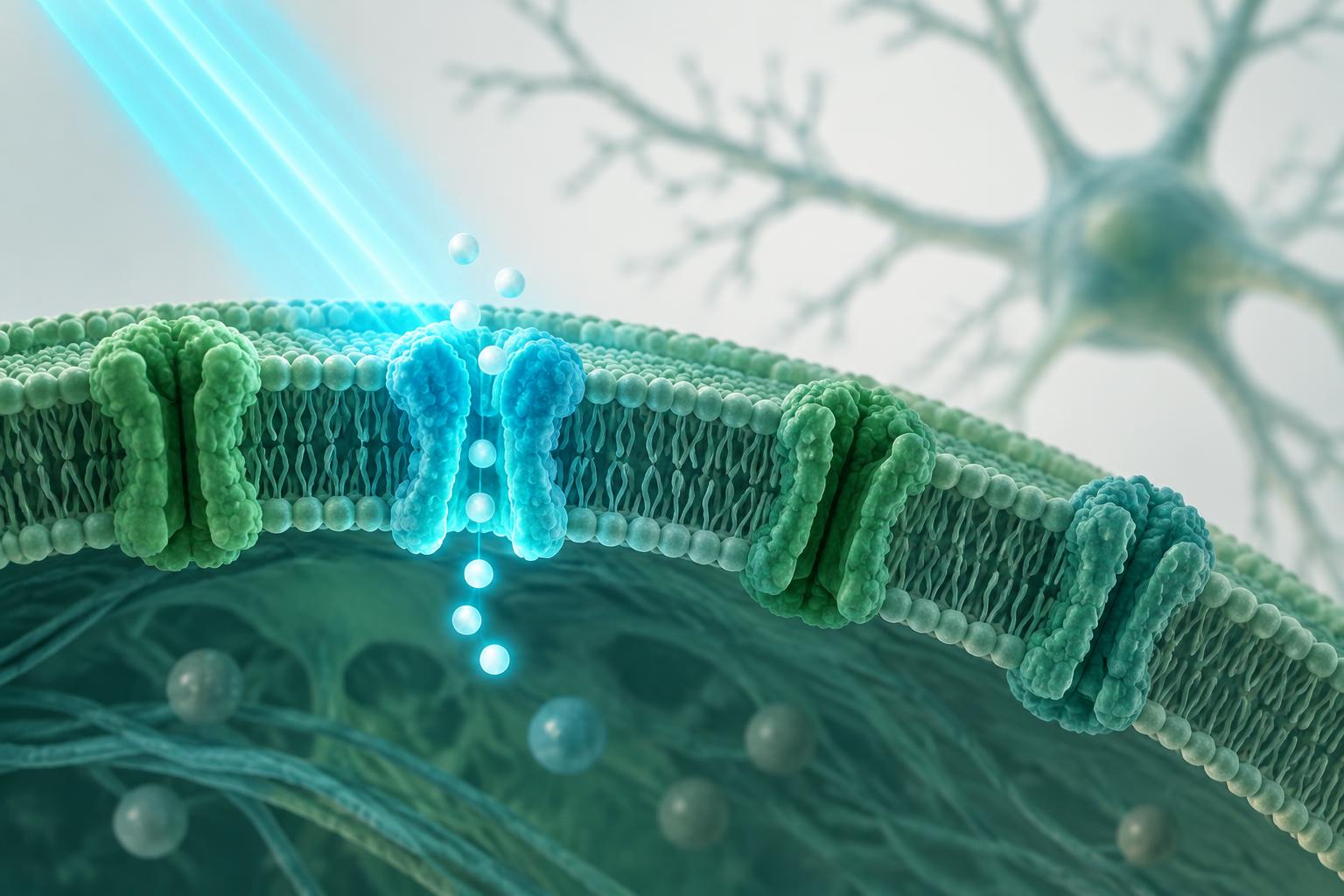
Target the cells
Genetic methods deliver instructions for a light-sensitive protein to selected cells.
Deliver the light
A suitable wavelength activates the protein, often using specialized optical equipment.
Observe the change
Researchers measure effects on activity, behavior or a disease-related process.
This approach is especially useful for testing cause and effect. If changing a particular pathway changes a behavior, that supplies stronger evidence of its role than a recording that merely shows the pathway was active. Even then, the result depends on the model and experiment; it does not explain every aspect of a human condition.
What can it reveal about brain disorders?
Brain disorders involve more than a single “broken” neuron. They can reflect changes in connected networks, and people with the same diagnosis may have different underlying biology. Optogenetics lets scientists break a broad question into smaller, testable circuit questions.
Anxiety: separating neighboring pathways
A 2011 study by Kay Tye and colleagues used optogenetics to investigate connections within amygdala circuitry. Manipulating a specific pathway altered anxiety-related behavior in mice. The finding illustrates why targeting a connection can reveal something that broadly stimulating a brain region cannot. It should not be read as a demonstration that human anxiety has been cured with light. [3]
Parkinson’s disease: understanding movement circuits
In a 2010 study, Alexxai Kravitz and colleagues—including Deisseroth—used optogenetics to examine two basal ganglia pathways. Activating different pathways had different effects on movement, and direct-pathway activation improved selected motor deficits in a mouse model of Parkinson’s disease. These results sharpened understanding of movement control; they did not establish a routine optogenetic therapy for patients. [4]
Epilepsy: interrupting seizures at the right moment
Esther Krook-Magnuson and colleagues reported a closed-loop system that detected and interrupted spontaneous seizures in a mouse model of temporal lobe epilepsy. Light was delivered in response to detected seizure activity, rather than continuously. The study suggests how precision in both timing and cell selection might matter for future interventions. This was work by a wider research community, not a clinical treatment offered by Deisseroth. [5]
Depression and other psychiatric conditions
Optogenetic experiments also investigate circuits associated with reward, motivation, stress and social behavior. These are relevant to psychiatric research, but a behavior measured in a rodent is not a complete model of depression or another human diagnosis. A useful study can reveal a mechanism without predicting which treatment will help an individual person. [6]
| Research area | What studies investigate | Important boundary |
|---|---|---|
| Anxiety | Amygdala pathways and anxiety-related behavior | Animal behavior is not a human diagnosis |
| Parkinson’s | Movement and basal ganglia circuitry | Model results are not a patient treatment |
| Epilepsy | On-demand control of seizure activity | Safety and translation need further study |
| Depression | Reward, motivation and related circuits | No single circuit explains every case |
From a research tool to a treatment
The distance between an elegant experiment and a safe treatment is substantial. Researchers need ways to deliver genetic material reliably, target the intended cells and supply light without unacceptable tissue damage. They also need to assess immune responses, long-term protein expression and whether changes in one circuit have unintended effects elsewhere. [2]
A larger and more complex human brain adds practical obstacles to light delivery. Most importantly, a result needs to translate into a meaningful benefit for patients—not only a change in a laboratory measurement. Optogenetics is therefore best understood here as a powerful research approach, rather than a ready-made solution for anxiety, depression, Parkinson’s disease or epilepsy.
It is also different from a nutritional product. A capsule does not introduce precisely targeted opsins and optical control into brain circuits. Nothing in the studies cited here establishes the safety or effectiveness of a commercial supplement, or shows that Deisseroth endorses one.
Common questions
Is optogenetics the same as light therapy?
No. Optogenetics requires genetically introduced light-sensitive proteins in targeted cells. Bright-light therapy and other light-based procedures use different mechanisms and should not be described as interchangeable.
Did Karl Deisseroth invent optogenetics alone?
No. He is a pioneering contributor, but the field developed through collaborative work on microbial light-sensitive proteins, genetic targeting and optical control. The authors of the landmark 2005 study are listed in the references.
Can optogenetics cure brain disorders?
The research cited here demonstrates specific effects in experimental models, not a general cure for human brain disorders. Decisions about treatment should rely on clinical evidence and a qualified clinician’s assessment.
Can a brain-health supplement work through optogenetics?
A supplement is not an optogenetic intervention. The studies discussed here do not validate a supplement’s ingredients, health claims or association with Karl Deisseroth.
Sources & further reading
- Boyden et al. (2005). Millisecond-timescale, genetically targeted optical control of neural activity. Nature Neuroscience.
- Optogenetics methods and applications review. Full text available through PubMed Central.
- Tye et al. (2011). Amygdala circuitry mediating reversible and bidirectional control of anxiety. Nature.
- Kravitz et al. (2010). Regulation of parkinsonian motor behaviours by optogenetic control of basal ganglia circuitry. Nature.
- Krook-Magnuson et al. (2013). On-demand optogenetic control of spontaneous seizures in temporal lobe epilepsy. Nature Communications.
- Tye & Deisseroth (2012). Optogenetic investigation of neural circuits underlying brain disease in animal models. Nature Reviews Neuroscience.
Editorial disclosure: This is an AI-assisted educational article, not a clinical review or medical advice. Images are generated conceptual illustrations. Commercial links are separate from the cited evidence; no scientist endorsement or verified purchase reviews are claimed. Consult a qualified healthcare professional about symptoms or treatment.